








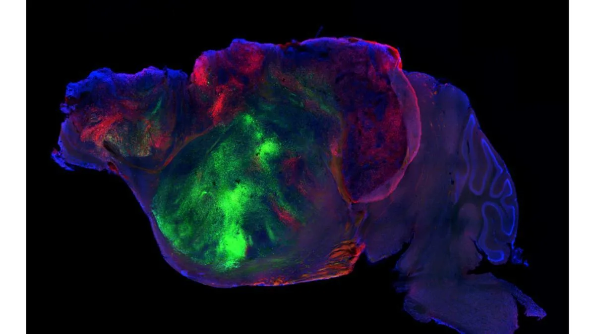
Human brain organoids implanted in mice have been developed and have formed neural networks of the cortex, the layer of gray matter that controls functions such as cognition, language, and decision-making. The breakthrough, presented today in the journal Nature, improves the ability to grow human brain tissue for medical research.
Read more The heavy rains are already falling in Catalonia and leaving a strong hailstorm in Girona
The authors of the work, from Stanford University in California, hope it will accelerate the study of neurodevelopmental disorders and the search for new therapies for neuropsychiatric diseases. Among the first to potentially benefit from this line of research are cerebral palsy, frontotemporal dementia, and autism spectrum disorders.
“Neuropsychiatry has fewer therapies than any other branch of medicine,” explained Sergiu Pasca, director of the research, at a press conference on Monday. Partly because the brain is more complex than other organs and partly because it is less accessible. “Our goal is to make the brain accessible to investigate therapies.”
Human neurons implanted on the brains of mice occupy half of the animals’ cranial cavity
Pasca began his line of research by creating brain organoids in the laboratory from human stem cells, but in vitro cultures did not reflect the complexity of the brain in vivo nor did they allow the study of behavior. He continued implanting human brain organoids in rats, but found that the rodents’ neurons expanded faster than the human ones and left them no space to grow, which is explained because human development is slower than that of rats.
In the new research, Pasca has genetically modified mice so that they do not develop the cerebral cortex or hippocampus, so that human neurons have more space to grow. The animals are healthy and behave similarly to normal mice, except that they move more carefully (indicating poorer motor coordination) and have worse memory (because they lack a hippocampus).
“We have taken extraordinary ethical oversight measures to care for the welfare of the animals,” Pasca states. “The university [Stanford] has created an ethics committee external to the university itself” to oversee the project. The researcher organized a bioethics congress last year on the use of human stem cells and the implantation of organoids in animals. “A predominant argument questioned the ethics of not doing this research considering the suffering of hundreds of millions of people affected by neurological disorders that are currently incurable,” Pasca recalls.
Despite having half of the brain formed by human neurons, mice do not develop our species’ abilities
The researchers implanted about four million human neurons in each mouse. As expected, the organoids developed and occupied the space left free in the cranial cavity. The neurons of the organoids differentiated into different types of human cortex cells, organized into active neural networks, integrated with the mice’s nervous system, and formed connections with the spinal cord. Once development was complete, almost half of the volume of the mice’s brains was made up of human neurons.
Read more Follow here the meeting of Enric Juliana with the subscribers of La Vanguardia, live
However, the organoids did not form glial cells—the other major type of cell present in the brain, alongside neurons—and did not reproduce the full complexity of the cortex. “There is no improvement” in the mice’s abilities, Pasca clarified at the press conference.
“Neurons do not have consciousness on their own,” recalls Óscar Marín, director of the Center for Developmental Neurobiology at King’s College London, who did not participate in the research but is well acquainted with Pasca’s work. “Just as having a pig heart valve does not make a person part pig, here the mouse remains a mouse.”
According to Marín, the research is “a technical advance” that “allows us to ask how human cortical neurons develop” and that “could be a good platform to check if a future drug or gene therapy modifies neuron behavior.”
In one of the most promising results of the research, the organoids have formed spindle neurons in the mice. This is a rare type of neuron that represents only one in every 90,000 neurons in the human cortex. They are found in brain regions involved in sociability and decision-making and are severely affected in frontotemporal dementia (the type of dementia Bruce Willis has). Until now, it had not been possible to create them in the laboratory. Obtaining them in mice from human cells opens the way to investigate why they deteriorate and to test therapies that protect them, Pasca highlights.
On the other hand, the researchers subjected mice with developing brains to a hypoxia situation to simulate the lack of oxygen that can cause cerebral palsy. The mice developed movement disorders characteristic of the condition. “We do not lack ideas to treat [neurodevelopmental diseases]. We lack a platform to test them,” Pasca states. Mice with human organoids “could be used to test therapies for cerebral palsy.”
The expert’s opinion
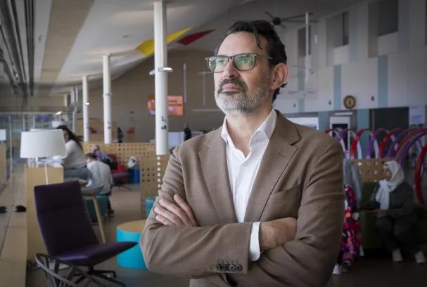
Óscar Marín: “It could be a good platform to test therapies”
Neuroscientist Óscar Marín, director of the Center for Developmental Neurobiology at King’s College London, positively evaluates Sergiu Pasca’s research with human brain organoids implanted in mice, although he warns that it is far from reflecting the complexity of the human brain.
What does this research contribute?
It is a real technical advance. Pasca’s laboratory demonstrated a few years ago that human cortical organoids transplanted into a rodent’s brain survive and mature. The obstacle was competition. Human neurons develop much more slowly than mouse neurons, so they arrive late and lose out when connections are established. In this new work, they eliminate that competition completely by genetically removing the mouse’s excitatory cortical neurons before birth. The human tissue then occupies most of the empty space, extends long-range projections reaching the spinal cord, and generates cell types very difficult to obtain in culture.
What does it allow that was not possible before?
It allows us to ask how human neurons normally connect and how much of their development is due to an intrinsic molecular program of the cell and how much is imposed by the environment. The important caveat is that the connections studied are still between human and mouse neurons.
For which disorders could it be most useful?
For those affecting the development of the cerebral cortex. The authors show an example: when the transplant is deprived of oxygen, a cellular and behavioral response occurs in the mice, offering a plausible way to study perinatal hypoxia and cerebral palsy.
Any other example?
Genetic neurodevelopmental disorders fit naturally, because cells carrying a patient’s mutation can be cultured alongside control neurons and compared in the same environment. But I think it is important to point out a limitation. The transplant contains very few human interneurons, and inhibition alteration is central in autism, epilepsy, and schizophrenia. Until human interneurons can be added, the model only captures half of the neuronal components of the cortical circuit.
What could be the short-term applications?
The most immediate is that it allows observation of how human cortical neurons develop, molecularly and electrically, in a much more realistic environment than a culture. Also, it could be a good platform to check if a drug or gene therapy modifies human neuron behavior.
Read more The Government activates the negotiation of the 2027 budgets


